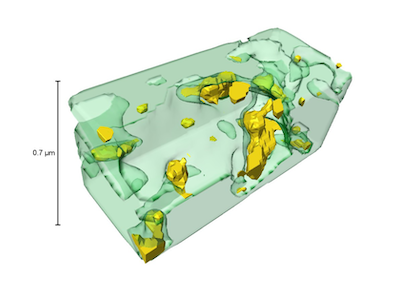
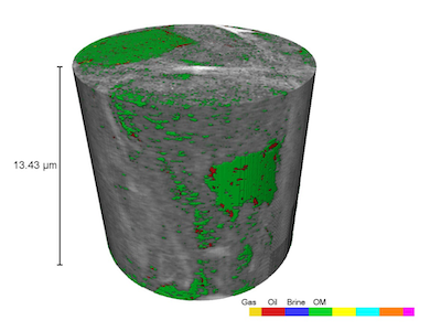
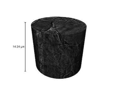
The high-resolutions obtained with this instrument allow researchers to investigate petrophysical properties along with fluid flow behavior at the nanometer scale. To obtain critical rock properties such as porosity, pore structure and pore-size distribution as well as the necessary flow parameters, including but not limited to, permeability and three-dimensional fluid occupancy maps, non-destructive X-ray computed tomography is used to probe the specimen at nanometer resolution. Samples used in the nano-CT are typically less than 20 μm in diameter and are prepared using focused ion beam milling (FIB) in a scanning electron microscope (SEM). During nano-CT imaging, a stack of 2D images of the sample is generated by passing X-rays through the sample at different orientations. The images are then put into image-processing software used to visualize the sample and establish a 3D view of the pore structure and fluid distribution throughout the sample.
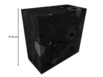
The Ultra XRM L-200 possesses two levels of resolution which allow the sample to be scanned in two different imaging modes. Large Field of View (LFOV) produces 150 nm resolution with a maximum field of view of 60 μm. High Resolution (HRES) settings hone the X-ray beam to produce pixel resolutions of 16 nm. These two modes of scanning allow users to characterize a wide range of samples including shale samples which possess many sub-micron pores that cannot be analyzed using a micro-CT scanner. The nano-CT machine can significantly improve the understanding of the pore-scale physics involved in multi-phase flow and transport phenomenon, not only in tight rock samples but also in other nanoporous materials. The Helios FIB-SEM instruments not only enable precision sample preparation for the nano-CT but also allow for high resolution imaging using cutting-edge 3D FIB imaging technology. By using a high energy ion beam, the FIB-SEM is able to ablate away thin layers of a sample. Between milling sequences, the electron beam is used to image the milled area at spatial resolutions of up to 2 nm. By subsequent milling and imaging, a three dimensional image of the sample can be constructed. These 3D images, when coupled with the results of the nano-CT allow for a better picture of the structure and connectivity of a wide variety of nanoporous materials including samples from unconventional oil and gas reservoirs.
GALLERY