This advanced technique is currently in use within the petroleum engineering and geoscience community and is used to investigate various aspects of multiphase flow phenomena in porous media. These state-of-the-art techniques are currently in play at the Center of Innovation for Flow Through Porous Media (COIFPM), enabling researchers to overcome the complex challenges in enhanced oil and gas recovery projects while developing deeper insights into the fundamental science of fluid flow in porous media.
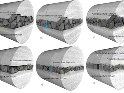
At the COIFPM, several conventional and helical micro-CT scanners are at work to image complex flow scenarios that have never been studied. Using this world class equipment enables visualization of the fluid occupancy in rocks with a spatial resolution of less than a micron. This capability has opened a host of opportunities to better understand the complicated capillary interactions and fundamental science at the pore level. The current capabilities are improving the current understanding of fluid transport in complex, artificially and naturally occurring porous media. These micro-CT instruments have also been equipped and integrated with custom closed-loop core-flooding systems. The system uses a state-of-the-art core holder designed to handle two- and three-phase fluid injections while including the effects of confining pressure, elevated pore pressures of up to 5000 psi, and a dynamic range of temperatures in order to accurately represent a wide range of real-world conditions. The core holder is mounted inside the micro-CT scanner allowing a 5-10 millimeter diameter core sample to be scanned while up to three fluids are injected simultaneously into the core.
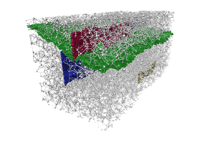
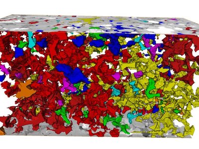
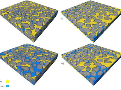
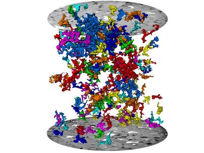
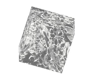
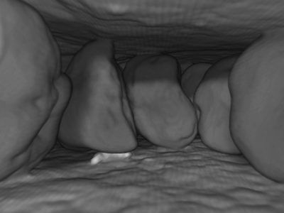
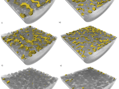
The tomograms (raw image from CT scanners) are analyzed using innovative image processing methodologies. Such methods allow for three-dimensional visualization of the pore space within the reservoir rock under different experimental conditions. Pore-fluid configurations and saturation profiles can also be obtained by distinguishing different fluid phases in CT images using a modified histogram thresholding technique. This, in turn, sheds light on the underlying displacement physics at the pore scale level. Such understanding is further used to enhance algorithms that model multiphase flow through porous media.
GALLERY